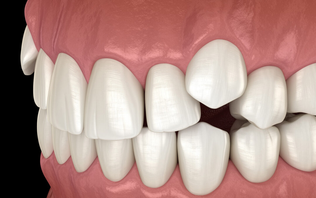
So, what is an impacted canine? An impacted canine is a permanent “eye tooth” that doesn’t erupt into the mouth on time or in the correct position. Because upper canines help guide your bite, support facial aesthetics, and protect other teeth during chewing, identifying and treating impaction early is important for long-term oral health. Below is an overview of causes, how an impacted canine is diagnosed, and the orthodontic and surgical treatments commonly used.
What Is an Impacted Tooth?
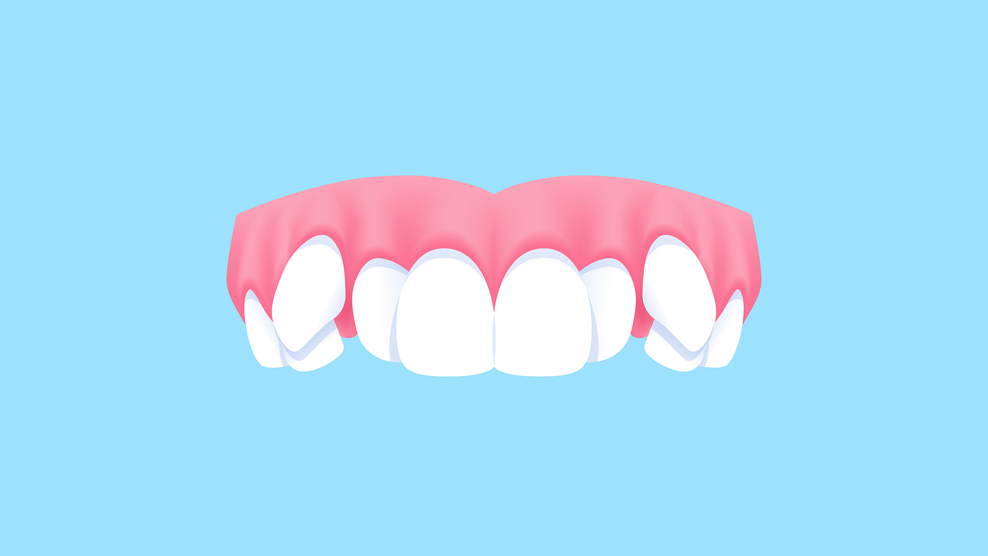
An impacted tooth is a permanent tooth that fails to fully emerge into its normal spot in the dental arch. Although any tooth can be impacted, an impacted maxillary canine (upper canine) is the second most frequently impacted after wisdom teeth. Lower (mandibular) canine impaction is less common but can still affect function and appearance. If you are wondering what an impacted canine is in practical terms, it is a canine that remains trapped in the jaw or palate and needs guidance or surgical assistance to reach its proper position.
Impaction is different from delayed eruption, in which a tooth is late but still follows a normal path, and from ectopic or partial eruption, where a tooth breaks through in an abnormal direction or location. With impaction, a barrier or an altered path of eruption prevents the tooth from coming in without assistance. This is especially relevant for an impacted maxillary canine, which often deviates toward the palate.
Possible signs include a noticeable delay compared with the opposite side, a baby canine that remains longer than expected, asymmetry in the smile, swelling or tenderness above the canine area, spacing or crowding in the upper front teeth, and occasional pain with biting or chewing. Sometimes there are no symptoms, and the issue is found on routine dental X-rays.
Causes and Risk Factors for Impacted Canines
Multiple factors can increase the likelihood of an impacted canine:
- Insufficient space in the upper jaw due to crowding
- Unfavorable tooth angulation or a blocked eruption path
- Retained primary (baby) canine that doesn’t shed on time
- Dental anomalies, such as peg-shaped lateral incisors, that alter normal guidance
Genetic influences can also contribute. A family history of impacted teeth, variations in tooth size or shape, and extra (supernumerary) teeth may disrupt normal eruption. Certain developmental conditions can affect timing and tooth movement, increasing the chance of an impacted maxillary canine.
Environmental and behavioral factors include dental trauma to baby teeth, premature loss of primary teeth that removes natural guidance, and oral habits that influence arch development. Scar tissue, cysts, or other pathology along the eruption path can also interfere and lead to an impacted canine.
Diagnosis and Early Recognition
Early recognition generally starts with a clinical exam. Upper canines typically erupt between ages 11 and 12. If a baby canine is still present beyond this timeframe or there is a clear right–left difference, an orthodontic evaluation is advisable. Additional signs, such as a bulge high in the gum over the canine region or the absence of a palpable canine “bump” by ages 10 to 11, may prompt imaging and referral.
Imaging helps determine the tooth’s position and its relationship to neighboring roots:
- Panoramic X-ray: Provides an overview of tooth development, angulation, and obstacles such as extra teeth.
- Periapical radiographs: Offer detailed, localized views of the root and surrounding bone.
- Cone beam CT (CBCT): Supplies 3D information about exact location and proximity to adjacent roots, aiding precise treatment planning and reducing risks.
Detecting an impacted canine early often allows simpler, more predictable treatment. Creating space and removing impediments before canine roots fully develop can guide the tooth into place, sometimes avoiding impacted canine surgery or later extraction.
Treatment Options: Exposure, Orthodontics, and Surgery
When an impacted canine is favorably positioned, conservative measures may be effective. Your orthodontist may monitor eruption with periodic imaging, extract a retained baby canine to clear the path, and use orthodontic appliances to create or maintain space. In selected cases, timely space creation alone allows the canine to erupt naturally, particularly for an impacted maxillary canine detected early.
If the tooth does not come in on its own, exposure and bonding is a common next step. An oral surgeon or periodontist makes a small opening in the gum (and sometimes bone) to uncover the canine and bonds a tiny bracket or attachment to it. The orthodontist then applies gentle traction using braces or clear aligners to guide the canine into its proper position. Treatment length varies from several months to more than a year, depending on the tooth’s location and response to movement. Typical short-term effects include gum soreness, minor bleeding, and swelling; irritation to adjacent roots is uncommon. Most patients resume normal activities within a day or two, with instructions for hygiene and comfort.
When the canine is severely malpositioned, fused to the bone (ankylosed), or threatens the health of adjacent teeth, surgical removal may be recommended. In these cases, the orthodontist and oral surgeon coordinate a plan to maintain bite function and appearance. Options may include closing the space orthodontically, redistributing space for a future dental implant once growth is complete, or reshaping adjacent teeth to mimic the canine. If exposure and traction are not feasible, impacted canine surgery can involve extraction and careful space management to preserve aesthetics and function.
A team-based approach tailors care to your age, stage of dental development, and smile goals. Whether treatment involves orthodontic traction after exposure or impacted canine surgery, early diagnosis and coordinated planning help protect neighboring teeth, support facial harmony, and achieve a stable bite.